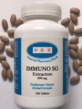
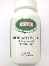
Research of Triptolide (Leigongteng) on Cancer:
Target and molecular mechanism of Triptolide in cancer therapy
Cuicui Meng, Hongcheng Zhu, Hongmei Song, Zhongming Wang,, Guanhong Huang, Defang Li, Zhaoming Ma, Jianhua Ma, QinQin, Xinchen Sun, and Jianxin Ma.
Chinese Juornal of Cancer Research, 2014 v.26(5); 2014 Oct. PCM US National Library of Medicine. National Institute of Health PMC4220249
Introduction:
Triptolide (TPL/TL), which is also called tripterygium wilfordii lactone alcohol or tripterygium wilfordii lactone, is extracted from tripterygium wilfordii celastraceae plants. TPL contains three epoxy groups of diterpene lactone compounds, and it has anti-tumor, anti-inflammatory and immunosuppressive activities. TPL exerts proapoptotic and anti-proliferative effects on tumor cell lines in vitro, and reduces tumor size or restricts tumor growth in vivo. Furthermore, TPL sensitizes tumor cells to other therapeutic methods, such as chemoradiation. The synergistic effects of TPL with other chemotherapeutic agents showed efficacy in preclinical animal models. This review summarizes the targets and molecular mechanisms of TPL in cancer therapy.
Triptolide (TPL) inhibits tumor cell proliferation:
Disorder of cell cycle regulation plays a significant role in the occurrence and development of tumors. The completion of the cell cycle is under the control of the serine/threonine kinase cyclin dependent kinase (CDK) family. The cell cycle can be completed when CDKs are activated. Periodic proteins (cyclins) positively regulate the cell cycle, and inhibitors of CDKs reverse regulation. Cell proliferation is out of control when CDK inhibitors are inhibited or cyclins are overexpressed, which leads to the occurrence of tumors. p21 is an inhibitor of CDKs. The expression of p21 inhibits the formation of all cyclin-CDK complexes. Inhibiting the expression of cell cycle proteins and CDK2 phosphorylation leads to cell cycle arrest, which inhibits cell proliferation. TPL up-regulates p27 and p21 expression, but down-regulation of CDC25A and cyclinA expression leads to S phase arrest. TPL inhibits the proliferation of colon cancer cells via inhibition of messenger ribonucleic acid (mRNA) expression, which affects cyclin expression. CyclinB promotes the G2-M transition. CyclinA and CyclinD are the regulatory proteins of G1-S. TPL down-regulates the expression of cyclinA, cyclinB, cyclinC, and cyclinD, which causes cell cycle arrest and inhibits cell proliferation
Triptolide tumor cell apoptosis:
TPL activates signaling pathways of caspases.There are two signaling pathways of apoptosis. One pathway is the death receptor signaling pathway, which combines cell membrane receptors with ligands and leads to the activation of apoptosis precursors to induce cell apoptosis. Another pathway is the signaling pathway of mitochondria, which induces apoptosis factors, damages mitochondrial structure and function, promotes apoptosis molecules from mitochondria, releases cytochrome into the cytoplasm, and affects a series of biological characteristics, resulting in cell apoptosis. TPL-induced apoptosis occurs by the release of cytochrome c and activation of caspase-3, and it has been found to be associated with the down-regulation of Bcl-2 expression and the up-regulation of Bax expression. TPL induces apoptosis by regulating apoptosis-related protein expression, such as the regulation of caspase-3, caspase-9, poly adenosine diphosphate ribose polymerase (PARP), and Bcl-2 activation. TPL up-regulates caspase-3/9 expression and down-regulates Bcl-2 expression without changing Bax levels. TPL also down-regulates Mcl-1 mRNA and protein levels. Furthermore, TPL reduces Mcl-1 protein, which correlates with caspase activation and induces apoptosis. TPL inhibits Mcl-1 synthesis at the mRNA transcription level, and the underlying molecular mechanism is associated with inhibition of RNA polymerase II carboxyl-terminal domain (CTD) phosphorylation. TPL down-regulates the expression of the anti-apoptotic proteins Bcl-2 and MDR1 at the gene and protein level, and it increases the expression of pro-apoptotic proteins, such as Fas and Bax (9). TPL induces endoplasmic reticulum (ER) stress via the PERK/eIF2α pathway. TPL induces extracellular signal-regulated kinase (ERK) activation via the regulation of the expression of the Bcl-2 protein family, but it is not involved in the down-regulation of Bcl-xL expression.
Triptolide inhibits heat shock protein (HSP) expression:
The family of HSPs, including HSP27, HSP70, and HSP90, is a group of stress proteins. These small-molecule chaperones participate in the regulation of apoptosis, and they are involved in carcinogenesis. Evidence shows that HSP27 plays an important role in drug resistance, and it is highly expressed in drug-resistant cancers, including colon cancer, gastric cancer, ovarian cancer, and lymphoma. Significant elevation of HSP27 expression in aggressive androgen-insensitive cell lines and malignant pancreatic cancer cell lines predicts a more aggressive cell type and poorer clinical outcome. These studies suggest that HSP27 should be considered as a potential target for chemotherapy-resistant carcinoma. TPL down-regulates HSP27 and HSP70 expression. TPL inhibits the promoter of the HSP70 gene, which down-regulates HSP27 and HSP70 expression. TPL inhibits the promoter of the HSP70 gene, which down-regulates heat shock gene expression, and induces more sensitive cells to a stress-induced cell death, which may be closely related to cancer therapy. TPL down-regulates HSP70 expression in pancreatic cancer. The mechanism of HSP70 expression dowm-regulation is that it changes the acetylglucosamine modification of Sp1 to cause inactivation, which leads to cell death.
Triptolide induces cell apoptosis through the NF-kB pathway:
NF-κB is cell nuclear factor that is involved in transcription regulation in the process of inflammation, stress, cell growth and proliferation. NF-κB promotes cell proliferation, inhibits cell apoptosis, and plays an important role in the process of tumor development. NF-κB is a heterologous dimer composed of p50 and p65. TPL inhibits the transactivation effect of the p65 subunit of NF-κB to inhibit NF-κB activity and promote cell apoptosis. TPL inhibits growth via apoptosis induction in THP-1 cells. The mechanism of apoptosis is through caspase activation and NF-κB inhibition. TPL indirectly affects NF-κB and suppresses NF-κB signaling mainly through the AKT/GSK3β/mTOR pathway. TPL induces apoptosis in ovarian cancer through the suppression of NF-κB expression to reduce human epidermalgrowth factor receptor-2 (HER2), which is downstream of the PI3K/Akt-signaling pathway. TPL inhibits complex I of the mitochondrial respiratory chain (MRC) in SKOV3 platinum-resistant human ovarian cancer cells. TPL down-regulates reactive oxygen species (ROS), inhibits nuclear factor NF-κB activation and decreases the anti-apoptotic proteins Bcl-2 and X-linked inhibitor of apoptosis protein (XIAP) (17). The induction of apoptosis by TPL in undifferentiated thyroid cancer cells is not through the p53 pathway but via the NF-κB pathway. Therefore, TPL may become a drug used in the treatment of p53-mutation-type thyroid cancers.
TPL enhanses apoptosis that is induced other mechanisms:
Deoxyribonucleic acid (DNA) rupture is the most important threat for cellular genome stability. Blockade or incomplete DNA repair can lead to chromosome replacement, misses or breaks (19). TPL induced DNA damage and suppressed DNA repair-associated gene expression (mRNA) in A375.S2 cells, which may underlie the TPL-mediated inhibition of cell proliferation. TPL modulates apoptosis in MCF-7 cells via the induction of liposomal membrane permeability, up-regulation of the expression of pro-apoptotic proteins, and the inhibition of cell proliferation in a time- and dose-dependent manner.
Triptolide inhibits tumor metastasisi:
The metastasis of solid tumors is the main cause of patient death. Vascular endothelial growth factor (VEGF) is an important angiogenesis factor that induces endothelial cell proliferation and migration and promotes angiogenesis. VEGF plays an important role in tumor growth and metastasis. VEGF combined with its receptors releases numerous growth factors and cytokines, enhances the permeability of blood vessels, causes plasma protein extravasation, and provides the substrate for the growth of tumor cells. VEGF also stimulates the proliferation and migration of vascular endothelial cells and promotes angiogenesis formation. TPL can be a promising antiangiogenic agent. The potent antiangiogenic action of TPL occurs via inhibition of angiogenic pathways mediated by Tie2 and vascular endothelial growth factor receptor (VEGFR)-2. TPL inhibits human pancreatic cancer cell proliferation in a concentration- and time-dependent manner and down-regulates VEGF expression in vitro. Furthermore, medium from TPL-treated PANC-1 cells inhibits the proliferation, tube formation, and migration of Human Umbilical Vein Endothelial Cells (HUVECs). Analysis of CD31 expression, which is a marker of tumor angiogenesis, further showed that TPL down-regulated blood vessel formation in previous experimental models.
Triptolide enhances the effect of chemotherapy:
TPL sensitizes several cancer cell lines to chemotherapy in vivo and in vitro. TPL highlights the synergistic anti-tumor effect in cells in combination with many cytotoxic drugs. The synergistic anti-tumor effect of TPL and cisplatin or 5-FU down-regulates cancer cell viability in liver cancer cell lines in vitro and in nude mice and induces higher levels of apoptosis compared to single treatments. Furthermore, cells treated with TPL plus cisplatin or 5-FU exhibit a marked production of intracellular ROS and caspase-3 activity, down-regulate Bcl-2 expression and up-regulate Bax expression. TPL sensitizes cells to carboplatin activity. Previous studies showed that combined-agent-treated groups almost stopped growing, and tumor weights in vivo were much lighter than with single-drug treatment. TPL in combination with sorafenib is superior to single drug treatment in inducing apoptosis and down-regulating viability via decreasing NF-κB activity. Tumor growth inhibition rates in combined-agent-treated groups in a nude mouse model are increased compared to single drug treatment. TPL combined with oxaliplatin (OXA) effectively inhibits proliferation in the colon cancer cell line SW480 and induces cell apoptosis. The mechanism partly involves the inhibition the expression of target genes in the cell cycle and nuclear translocation of β-catenin. Moreover, combined-agent-treated groups in a nude mouse model significantly suppressed tumor growth.TPL in combination with temozolomide (TMZ) significantly up-regulates the percentage of apoptotic cells in glioma-initiating cells via up-regulation of NF-κB transcriptional activity and increased expression of downstream genes.
The combination of TPL with non-cytotoxic drugs has synergistic effects in numerous types of cancer cells. The main mechanism of the TPL-enhancing apoptosis effect of dexamethasone is that TPL affects the PI3k/Akt/NF-kB pathway, mitogen-activated protein kinase (MAPK) signaling pathway, and Bcl-2 expression (29). The synergistic antitumor effect of TPL and iron deficiency anemia (IDA) in acute myelocytic leukemia (AML) cells is due to induction of ROS and the inhibition of the Nrf2 and aspirin in cervical cancer involves a reduction in cyclin E expression, up-regulation of Bax and P21 expression, the inhibition of cell proliferation and induction of cell apoptosis.
Triptolide enhances the effect of radiotherapy:
TPL improves radiosensitivity and has synergistic antitumor effects in vitro and in vivo. Cell survival in combined-agent-treated groups in pancreatic cancer cells is significantly suppressed, and the percentage of apoptotic cells is significantly up-regulated compared to single treatment. Moreover, tumor growth in a nude mouse model is significantly suppressed in combined-agent-treated groups. Immunohistochemistry and terminal-deoxynucleotidyl transferase mediated nick end labeling (TUNEL) of caspase-3 cleavage in tumor tissues showed similar evidence. TPL in combination with radiation has synergistic anti-tumor effects on the expression of XIAP and Mcl-1 proteins in oral cancer cells especially. Tumor size and volume of combined-agent-treated groups in a nude mouse model were decreased significantly compared to single treatment groups. Caspase-3 expression was up-regulated and XIAP protein expression was decreased in this model.
Chinese Juornal of Cancer Research, 2014 v.26(5); 2014 Oct. PCM US National Library of Medicine. National nstitute of Health PMC4220249
Triptolide sensitizes resistant cholangiocarcinoma cells to TRAIL-induced apoptosis.
Pnichakul T, Itachote P, Wongkajorsilp A, Sripa R, Sirisinha S.
Laboratory of Immunology, Chulabhom Research Institute, BangKok, Thailand.
BACKGROUND: Tumor necrosis factor-related apoptosis-inducing ligand (TRAIL/Apo2L) promotes apoptosis by binding to transmembrane receptors. It is known to induce apoptosis in a wide variety of cancer cells, but TRAIL-resistant cancers have also been documented. In this study, the relative resistance of human cholangiocarcinoma (CCA) cell lines against TRAIL-induced apoptosis is reported and the possible potential synergistic effect with triptolide, a diterpene triepoxide extracted from the Chinese herb Tripterygium wilfordii, in killing TRAIL-resistant CCA cells is investigated. MATERIALS AND METHODS: Six human CCA cell lines were treated with various concentrations of TRAIL and the resistant cells were identified and subsequently tested for their sensitivity to a combination of TRAIL and triptolide. The susceptibility and resistance of the cells were based on analysis of cytotoxic and apoptotic induction and expression of anti-apoptotic factors (Mcl-1 and cFLIP). RESULTS: The treatment of TRAIL induced a dose-dependent decrease in cell viability in 4 out of the 6 cell lines. A combination of TRAIL and triptolide enhanced cytotoxicity and apoptosis in these 2 resistant cell lines. The combined treatment enhanced activation of caspase-8 and its downstream signaling processes compared with the treatment with either one alone. CONCLUSION: The results presented show that human CCA cells were heterogeneous with respect to susceptibility to TRAIL-induced apoptosis. The combination of TRAIL and triptolide could enhance susceptibility to TRAIL-induced apoptotic killing in these TRAIL-resistant CCA cells, thus offering an alternative approach for the treatment of TRAIL-resistant cholangiocarcinoma.
PMID: 16475706 [PubMed - indexed for MEDLINE]
Triptolide induces caspase-dependent cell death mediated via the mitochondrial pathway in leukemic cells.
Carter B Z, Mak D H, Schober W D, McQueen T, Harris D, Estrov Z, Evans R L, Andreeff M.
Section of Molecular Hematology and Therapy, Department of Blood and Marrow Transplantation, The University of Texas, MD Anderson Cancer Center, Houston, 77030, USA.
Triptolide, a diterpenoid isolated from the Chinese herb Tripterygium wilfordii Hook.f, has shown antitumor activities in a broad range of solid tumors. Here, we examined its effects on leukemic cells and found that, at 100 nM or less, it potently induced apoptosis in various leukemic cell lines and primary acute myeloid leukemia (AML) blasts. We then attempted to identify its mechanisms of action. Triptolide induced caspase-dependent cell death accompanied by a significant decrease in XIAP levels. Forced XIAP overexpression attenuated triptolide-induced cell death. Triptolide also decreased Mcl-1 but not Bcl-2 and Bcl-X(L) levels. Bcl-2 overexpression suppressed triptolide-induced apoptosis. Further, triptolide induced loss of the mitochondrial membrane potential and cytochrome C release. Caspase-9 knock-out cells were resistant, while caspase-8-deficient cells were sensitive to triptolide, suggesting criticality of the mitochondrial but not the death receptor pathway for triptolide-induced apoptosis. Triptolide also enhanced cell death induced by other anticancer agents. Collectively, our results demonstrate that triptolide decreases XIAP and potently induces caspase-dependent apoptosis in leukemic cells mediated through the mitochondrial pathway at low nanomolar concentrations. The potent antileukemic activity of triptolide in vitro warrants further investigation of this compound for the treatment of leukemias and other malignancies.
PMID: 16556893 [PubMed - indexed for MEDLINE]
Protection effect of triptolide to liver injury in rats with severe acute pancreatitis.
Zhao Y F, Zhai W L, Zhang S J, Chen X P.
Hepatic Surgery Center, Tongji Hospital, Tongji Medical College, Huazhong University of Science and Technology, Wuhan 430030, China.
BACKGROUND: The high mortality of patients with severe acute pancreatitis (SAP) is due to multiorgan dysfunction. The mechanisms of SAP are still obscure. The aim of this study was to investigate the role of nuclear factor-kappa B (NF-kappaB) activation in rats with SAP associated with liver injury and the protection effect of triptolide against liver injury in rats with SAP. METHODS: Ninety Wistar rats were randomly divided into three groups (n=30 each group): severe acute pancreatitis (group P), treatment with triptolide (group T), and sham operation (group S). SAP models were induced by retrograde injection of 5% sodium taurocholate to the pancreatic duct. After the model was successfully established, no treatment was given to group P. In group T, triptolide (0.05 mg/ml) was injected intraperitoneally (0.2 mg/kg). In group S, the abdominal walls of rats were opened, sutured, but not treated. The rats were sacrificed after operation at 2, 6, and 12 hours, respectively. The serum levels of amylase (AMY), alanine aminotransferase (ALT), tumor necrosis factor-alpha (TNF-alpha) and interleukin-6 (IL-6) were determined at three time points (10 rats for each time point). Liver tissues were obtained to detect the activity of NF-kappaB and to observe their pathological changes with light and electron microscopes. RESULTS: The serum levels of AMY and ALT were higher in groups P and T than in group S. The serum AMY levels were significantly lower in group T than in group P at 12 hours after operation. The serum ALT levels were significantly lower in group T than in group P at 6, 12 hours after operation. At the three time points, the levels of TNF-alpha and IL-6 in groups P and T increased more significantly than in group S. In group T they were decreased more significantly than in group P at the three time points. In groups P and T, NF-kappaB activity in liver tissue increased more significantly than in group S at the three time points. The activity of NF-kappaB was higher in group P than in groups S and T at the three time points. Liver pathological damages were milder in group T than in group P under light and electron microscopes. CONCLUSIONS: NF-kappaB plays an important role in the pathogenesis of liver injury in rats with SAP. Triptolide can reduce pathological damage to the liver. Its mechanism is to inhibit the activity of NF-kappaB and to decrease the release of inflammatory mediators.
PMID: 16286273 [PubMed - indexed for MEDLINE]
Triptolide, a diterpenoid triepoxide, induces antitumor proliferation via activation of c-Jun NH2-terminal kinase 1 by decreasing phosphatidylinositol 3-kinase activity in human tumor cells.
Miyata Y, Sato T, Ito A.
Department of Biochemistry and Molecular Biology, Tokyo University of Pharmacy and Life Science, School of Pharmacy, Hachioji, Japan.
Triptolide, a diterpenoid triepoxide extracted from the Chinese herb Tripterygium wilfordii Hook f., exerts antitumorigenic actions against several tumor cells, but the intracellular target signal molecule(s) for this antitumorigenesis activity of triptolide remains to be identified. In the present study, we demonstrated that triptolide, in a dose-dependent manner, inhibited the proliferation of human fibrosarcoma HT-1080, human squamous carcinoma SAS, and human uterine cervical carcinoma SKG-II cells. In addition, triptolide was found to decrease phosphatidylinositol 3-kinase (PI3K) activity. A PI3K inhibitor, LY-294002, mimicked the triptolide-induced antiproliferative activity in HT-1080, SAS, and SKG-II cells. There was no change in the activity of Akt or protein kinase C (PKC), both of which are downstream effectors in the PI3K pathway. Furthermore, the phosphorylation of Ras, Raf, and mitogen-activated protein/extracellular signal-regulated kinase 1/2 was not modified in HT-1080 cells treated with triptolide. However, the phosphorylation of c-Jun NH(2)-terminal kinase 1 (JNK1) was found to increase in both triptolide- and LY-294002-treated cells. Furthermore, the triptolide-induced inhibition of HT-1080 cell proliferation was not observed by JNK1 siRNA-treatment. These results provide novel evidence that PI3K is a crucial target molecule in the antitumorigenic action of triptolide. They further suggest a possible triptolide-induced inhibitory signal for tumor cell proliferation that is initiated by the decrease in PI3K activity, which in turn leads to the augmentation of JNK1 phosphorylation via the Akt and/or PKC-independent pathway(s). Moreover, it is likely that the activation of JNK1 is required for the triptolide-induced inhibition of tumor proliferation.
PMID: 16176806 [PubMed - indexed for MEDLINE]
The effect of triptolide on CD4+ and CD8+ cells in Peyer's patch of SD rats with collagen induced arthritis.
Zhou J, Xioa C, Zhao L, Jia H, Zhao N, Lu C, Yang D, Tang JC, Chan A S, Lu A P.
Institute of Basic Theory, China Academy of Traditional Chinese Medicine, Beijing 100700, China.
Triptolide is a purified component from a traditional Chinese herb Tripterygium wilfordii Hook F. It has been shown to have anti-inflammatory and immunosuppressive activities by its inhibitory effect on T cells. But the effect of triptolide on Peyer's patch cells is unknown. Enteric mucosal immune system, including Peyer's patch, is regarded as one of the sites for inducing immunity tolerance, and this intolerance effect has been used to induce oral tolerance which can considerably reduce arthritis severity in several models of experimental polyarthritis and RA patients. In this study, we investigated the effect of triptolide on the Peyer's patch cells and peripheral lymphocytes in collagen induced arthritis (CIA) in rats. CIA in rat is a widely studied animal model of inflammatory polyarthritis with similarities to rheumatoid arthritis (RA). Our data show that triptolide could lower the arthritic scores and delay the onset of CIA. There are more Peyer's patches in triptolide treated rats than in control rats, while there is no difference in Peyer's patch numbers between CIA rats and triptolide treated rats. In the Peyer's patch, more CD4+ cells are observed in CIA rats, and the numbers of CD4+ cells in triptolide treated rats and control rats are similar. While more CD8+ cells are observed in triptolide treated rats, and the numbers of CD8+ cells in CIA rats and control rats are similar. In periphery, more CD4+ cells and less CD4+ cells in CIA rats and triptolide treated rats are respectively observed. Therefore, the regulation on Peyer's patch might explain some of the immunosuppressive activities of triptolide, and enteric immune response might be actively involved in CIA pathogenesis. It is suggested that the Peyer's patch is one of the primary targets of the immunosuppressive activity of triptolide.
PMID: 16399624 [PubMed - indexed for MEDLINE]
Triptolide down-regulates tumor necrosis factor-a and interferon-g-induced overexpression of monocyte chemoattractant protein-1 in human proximal tubular epithelial cells
Heng LI, Zhi-Hong LIU, Chun-Sun DAI, Dong LIU, Lei-Shi LI
Research Institute of Nephrology, Jinling Hospital, Nanjing University School of Medicine, Nanjing, China.
Objective: Triptolide is the major active component of Tripterygium wilfordii Hook. f., which has been used as an anti-inflammatory agent in traditional Chinese medicine for centuries. The mechanisms of the anti-inflammatory effects of triptolide in renal diseases are not well understood. Recent studies have shown that overexpression of monocyte chemoattractant protein-1 in renal tubular epithelial cells is associated with the inflammatory injuries of renal tubulointerstitium. In this study, we investigated the effect of triptolide on the overexpression of monocyte chemoattractant protein-1 in renal tubular epithelial cells in vitro.
Methods: Human proximal tubular epithelial cells were treated with gamma interferon (200mg/L) and tumor necrosis factor-alpha (20mg/L) or combined with different concentrates of triptolide (0.4, 2, 10mg/L) for 12 hours (for monocyte chemoattractant protein-1 mRNA measuring) or 24 hours (for monocyte chemoattractant protein-1 protein measuring). The expression of monocyte chemoattractant protein-1 mRNA was detected by using reverse transcriptase-polymerase chain reaction. The expression of monocyte chemoattractant protein-1 protein in the cells was measured by using flow cytometry. Monocyte chemoattractant protein-1 levels in the supernatant were measured by using the enzyme-linked immunosorbent assay method.
Results: After treatment with interferon-g and tumor necrosis factor-a, monocyte chemoattractant protein-1 mRNA and protein levels in human proximal tubular epithelial cells and monocyte chemoattractant protein-1 concentration in the supernatant were significantly increased. Triptolide (10mg/L) can significantly inhibit the overexpression of monocyte chemoattractant protein-1 mRNA and protein in human proximal tubular epithelial cells. The increase of monocyte chemoattractant protein-1 protein in the supernatant was also markedly inhibited by triptolide (2, 10mg/L).
Conclusions: The results of this study suggest that the inhibition of monocyte chemoattractant protein-1 overexpression in tubular epithelial cells may contribute to the anti-inflammatory effects of triptolide in renal diseases.
(Hong Kong J Nephrol 2002;4(1):29-32)
Pharmacological details about Tripterygium wilfordii (Lei Gong Teng):
Key words:Epithelial cells/immunology, Enzyme-linked immunosorbent assay, Kidney tubules/pathology, Nuclear factor-kappaB (NF-kB), Tripolide
Wilfordine; Wilforgine; Wilfordine; Wilforine; Wilfotrine; Wilforzine; Triptolide; Tipdiolide; Tripteroilide; (+)-medioresinol; (-)syringaresinol; Nubiletin; Tripdiolide; Triptolidenol; Hypolide; Triptonide; 16-hydroxytriptolide; Tripchlorlide; Triptriolide; Tripdiotolnide; Wilforlide A, B; 3-epikatonic acid; Cangoronine; Regelin; Salaspermic acid; Euonymine; Wilfordsine; b-sitosterol; Dulcitol; Evonymine; Polysaccarides; 1-epicatechin; Triptotriterpenonic acid A; Triptotriterpenoidal lactone A; Oleanane-9,12-dialkene-3-ketone; 16-hydroxy-19, 2O-epoxy-kaurane; 13, 14-epoxide 9, 11, 12-trihydroxytriptolide; 3-hydroxy-2-oxo-3-fridelen-20a-carboxylic acid; 3b-hydroxy-D:B-friedoolean-5:6-epoxy-29-oic acid; Triptofordins A, B, C-1, C-2, D-1, D-2, E, F-1, F-2, F-3, F-4.
Dosage:
decoction: 5-25g if the bark of the root and woody parts are removed, 10-12g if the bark of the root is removed. Decoction requires small fire, 1-2 hours.
Can also be made into syrup, if powder is used to make capsules, 0.5-1.5g each time, 3 times per day.
Caution:
Acute toxicity test on rats show that Lei Gong Teng cortex can induce significant pathologic change to cardiac muscles, forming multiple tiny myolysis focuses, causing renal tuber cell degeneration and necrosis, and lymphocyte disintegration. In rats, the organs most sensitive to the toxicity of Lei Gong Teng total saponins are those of the gastrointestinal system, the hematopoietic system, and the germinal epithelium of testis. Either prolonged use or overdose of Lei Gong Teng inhibits white blood cells and blood platelets in dogs, and can cause damages to the germinal epithelium of dogs, rats, and mice, inhibiting spermatogonium division and resulting in a decrease in, or complete disappearance of, various germ cells.
1. Exercise caution when administering to patients with heart, liver, or kidney problems, or with a low blood cell count.
2. Contraindication: Pregnancy.
3. Adverse effects: There have been reports of one case of Leigongteng overdose, and one case of Leigongteng poisoning resulting in renal failure.
Effects on the reproductive system:
Mice that have been fed Lei Gong Teng syrup for six months are found to have lowered SDH, LDH, and ATPase activity levels in their various spermatogenic cells in the convoluted tubule of testis, heightened ACP activity in the supporting cells, while the activity levels of SDH, ACP, AKP, and ATPase in Leydig's cells remain more or less unchanged (an exception is the activity of LDH, which is increased).Furthermore, experiments show that the monomer T9 (triptolidenol) of Tripterygii Wilfordii Polyglycosides (TWP) is a potent spermicide, with a potency 100 times that of TWP.
Immunoregulatory effects:
TWP has an inhibitory effect on both the activation and proliferation of T cells. Used to treat early-stage nephrotoxic serum nephritis of mice, TWP can significantly suppress spleen lymphocytes' production of IL-2, check the level of serum anti-kidney antibody, reduce urinary protein output, and ameliorate renal pathological changes. Moreover, experiments on TWP's immunoregulatory effects in rabbits of acute experimental serum syndrome show that it inhibits ConA-induced proliferation of T cells (and therefore the production of IL-2) and the forming of circulating immune complexes (CIC), and reduces the inflammatory cell infiltration and immune complex sedimentation in the glomerulus.
Suppressing graft-rejection:
Experiments on Lei Gong Teng's effects on heterogenous corneal transplant rejection reaction show that it can postpone the rejection reaction and significantly decrease the percentage of T helper lymphocytes in the peripheral blood. Similar effects are observed on small intestine transplant rejection reaction in pigs and lung transplant rejection reaction in rats.
Anti-inflammatory effects:
Lei Gong Teng has a significant inhibitory effect on both carrageenin-induced swelling and adjuvant arthritis in rats. It can also suppress the production of IL-1 by the peritoneal macrophage in AA rats and the production of hemolysin antibody in mice.
Anti-adjuvant arthritis effects:
Administered to rats of adjuvant arthritis for seven consecutive days, ethyl acetate-based extract of Lei Gong Teng can lower the subjects' maximum platelet aggregation rate. TWP can significantly decrease the levels of IL-1 and IL-6 in the serum of AA rats, decrease the level of inflammatory cell factors in the joint fluid, and significantly curb the peritoneal macrophage's and spleen lymphocytes' ability to secret cell factors.
Antineoplastic effects:
A new component that has an antineoplastic activity has been isolated from Lei Gong Teng extract. Administered by abdominal injections for 2-3 times, this new component of Lei Gong Teng can significantly prolong the survival time of mice of H22, S180, EAC, and mammary cancer. Fed to mice of S37 and of 3-MCA-induced experimental lung cancer, it can inhibit the growth of the respective tumors by 42% and 65.13%. At 10 or 20mg/ml, it can kill 95% of HL60 cells in 48 hours, and at 20 or 40mg/ml, it can kill 90% of Daudi cells in 48 hours. At 5.10mg/ml, it enhances the phagocytic function of isolated mouse peritoneal macrophage; at 20mg/ml or above, however, its action reverts to inhibition.
Miscellaneous effects:
1) TWP (TI) can significantly suppress glomerulate membrane cells'(GMC) ability to synthesize anion peroxide and hydrogen peroxide. It can also clear GMC synthesized active oxygen. 2) Lei Gong Teng can reduce urinary protein output in rats with adriamycin-induced nephrosis in rats, increase the level of serum total protein and decrease that of lipid peroxide, promoting the structural recovery of the glomerulate epithelial cells. 30 3) TWP (II) has a dose-dependent inhibitory effect on the in-vitro proliferation of mice's hematopoietic stem cells, with the 50% inhibition dose being 0.5mg/ml. Within 8 hours, this inhibitory effect is reversible. After 8 hours, however, it becomes irreversible. Contact


